Anatomy Trains faculty and AT dissections assistant teacher, Lauri Nemetz spent a month at the end of summer in Guben, Germany as part of the international volunteer team of dissectors from around the world for the next phase of the Fascial Net Plastination Project with the support of Anatomy Trains. Below is the guest blog post from Lauri about her experience. Enjoy!
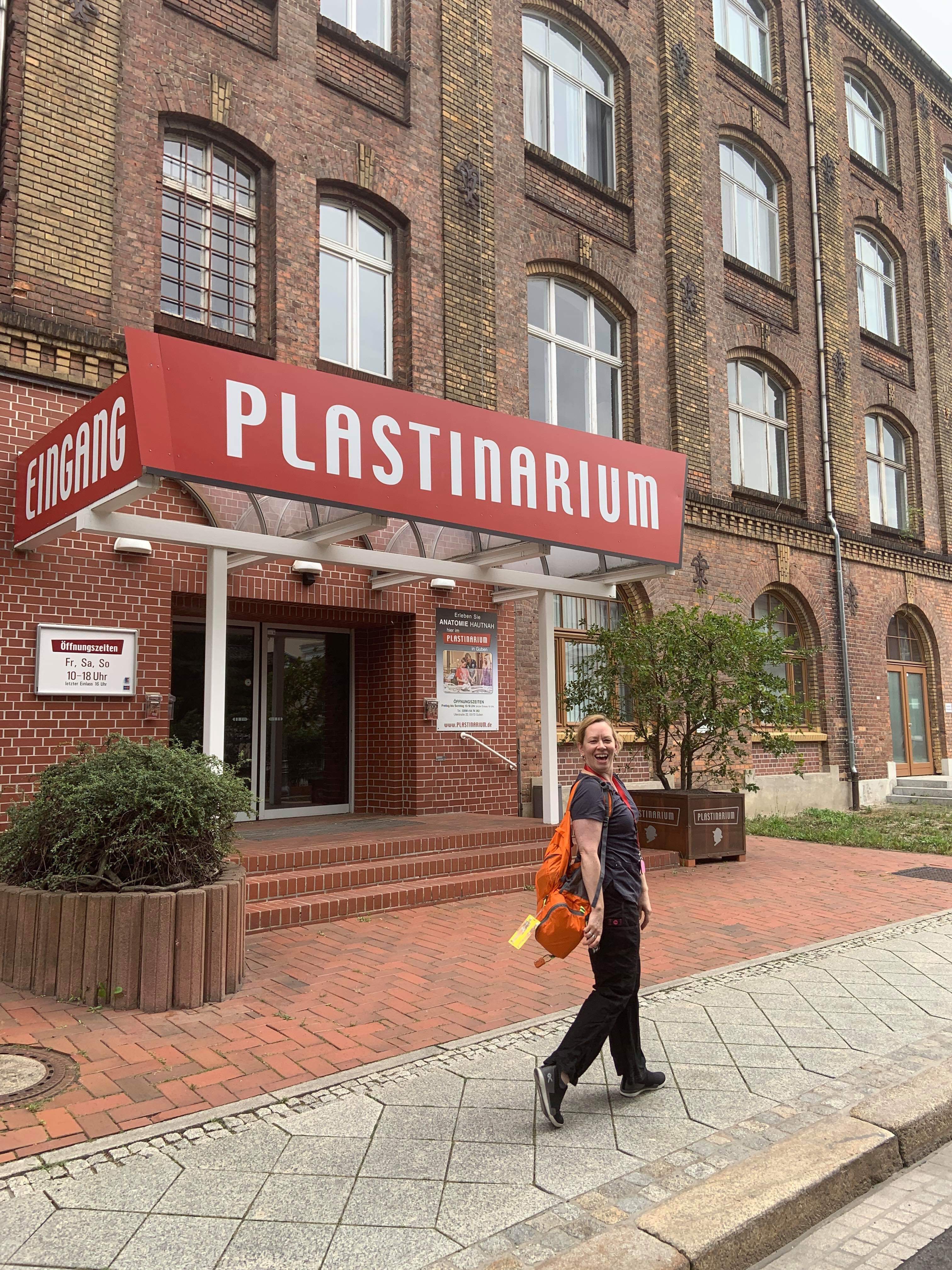
“Even the depths of things hold only surfaces” ~ Gunther von Hagens (quoted in placard at the Plastinarium, Guben, Germany)
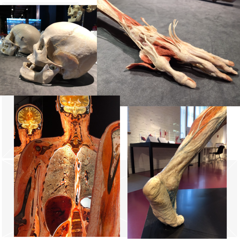
Over the years, I have been deep diving into the world of dissection and fascia and have recently returned from several weeks of intensive work as part of the next phase of the Fascial Net Plastination Project. This project began in January 2018 as a collaboration of the Fascia Research Society, Somatics Academy, the Plastinarium, and Body Worlds, to create the first human, fascial, plastinated specimens, which were exhibited in Berlin at the 5th International Fascia Research Congress. The current phase of the project is working to create the first fascial focused, full-body plastinate. If successful, it will debut at the 6th International Fascia Research Congress in the fall of 2021 in Montreal, Canada.
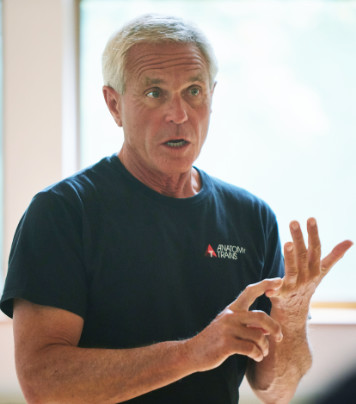
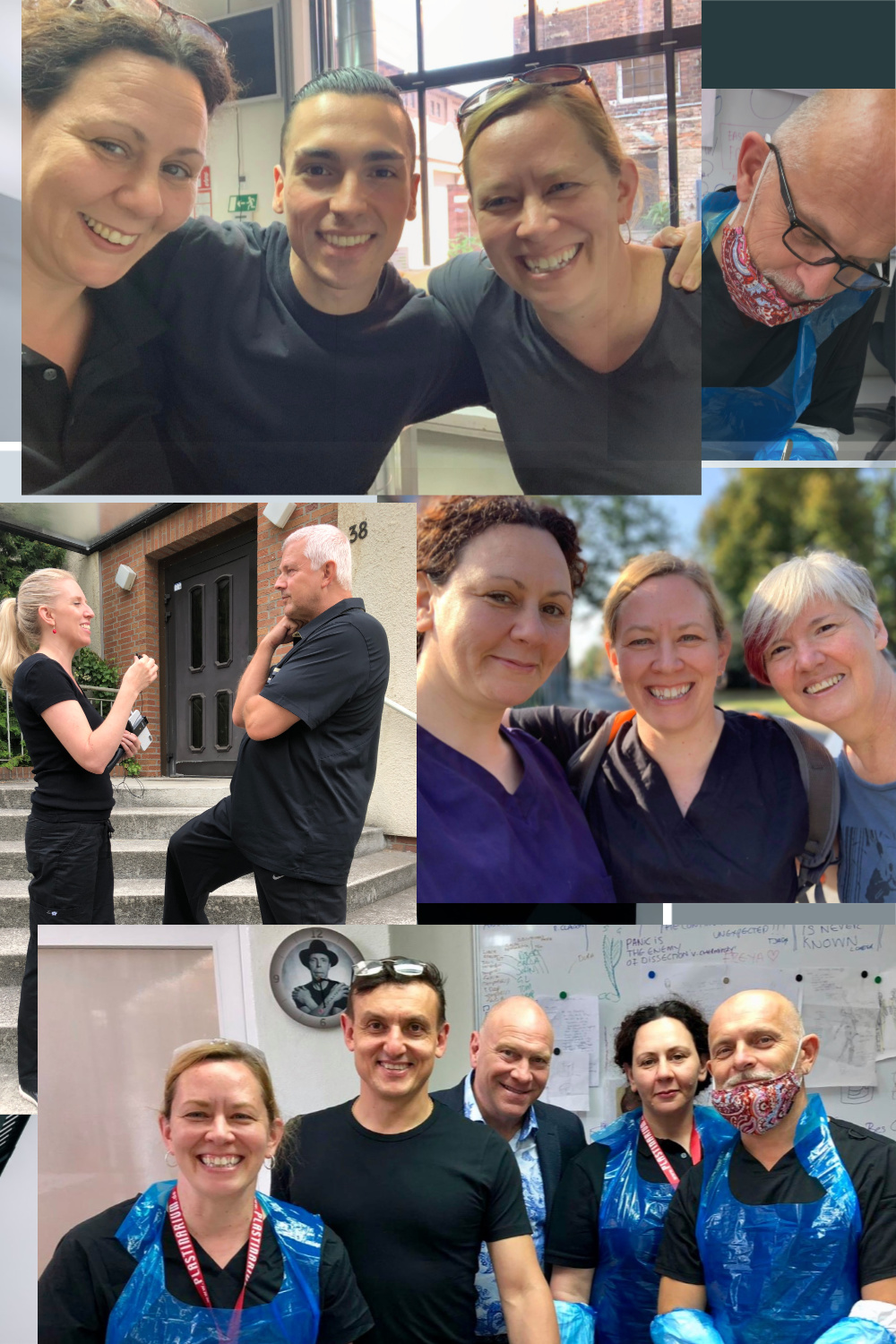
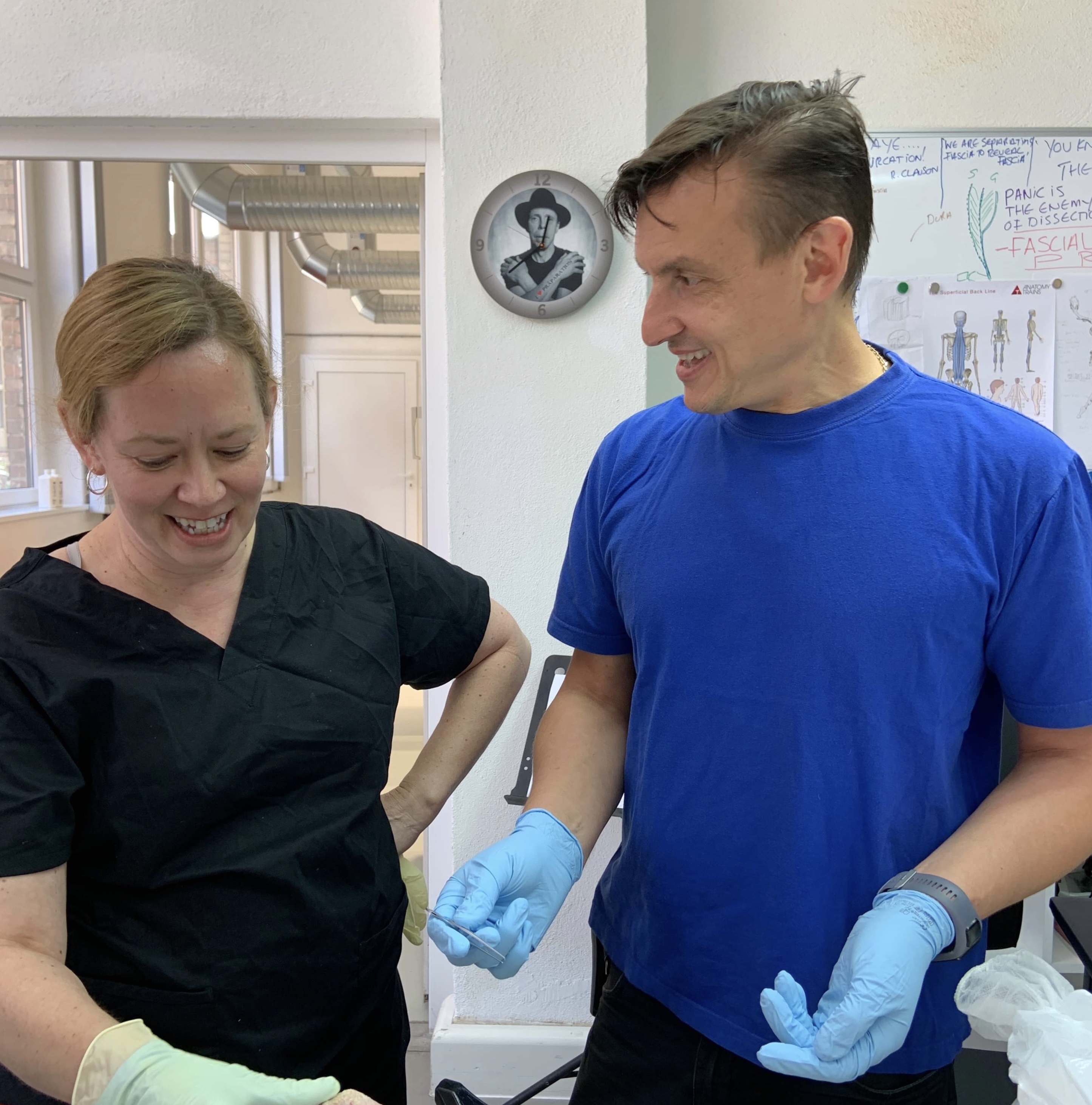
I spent a total of four weeks in Germany. It was a challenge to clear my schedule, but it was important to me to be a part of this dissection project, which has the potential to greatly impact the furthering of our understanding of fascia. The efforts of the entire international team have come together to work on a whole body form and to create more individual specimens for future exhibitions. During my time in the lab, we gathered in teams of two or three people, additionally supported by Plastinarium staff team member, Alexandro Navarro Valdivia, and overseen by Dr. Vladimir Chereminskiy, the Plastinarium Director of Anatomy and Plastination. Gary Carter has beautifully drawn the designs for this phase of the project, created with input from our expert fascial anatomy advisors and the FNPP team. The final plan is continually being revised due to challenges and opportunities we discover as things progress.
One of the first things I did upon my arrival was be a part of the first FNPP webinar, held on July 29th, about the project’s past, present, and future plans (scroll down under https://fasciaresearchsociety.org/online-lectures and search for Fascia Net Plastination Project). Organized by Fascia Research Society President, Anita Boser, the webinar was hosted by FNPP Communications Coordinator and team member Rachelle Clauson with project director Dr. Robert Schleip, FNPP Phase 3 Coordinator Gary Carter, Chief Photographer Stefan Westerback, and myself as expert dissector and team member. This particular group was also a part of the Exhibition Committee who produced “Fascia in a NEW LIGHT: The Exhibition” at the FRC5 in Berlin last November, which displayed the plastinates created during the first phase of the project. An audio and visual guide, recorded with the voices of FNPP team members and images of the specimens, is still available via the free Otocast app (for Apple or Android devices). In the app, search for ‘fascia’ and choose “Fascia In a NEW LIGHT” in English and German to view the exhibit from Berlin, Germany or choose the upcoming conference featuring the plastinates in Sao Paulo, Brazil to read and listen in Portuguese.
The nature of the plastination process is highly collaborative, and the interest in fascia as a body-wide network is somewhat new to the Plastinarium. We are all working together as we explore the techniques needed to properly preserve areas of fascial focus and still follow the required procedures for the plastination process to be successful all around. We are also working with preserved tissue, which has the advantage of withstanding the processes of dissection over a longer period of time than fresh tissue, but has the disadvantage of losing some of the finer qualities that unpreserved fascial tissues possess, particularly in terms of mobility.
As the weeks continued, it was quite exciting to have the staff ask us more and more questions about current definitions of fascia versus traditional, clinical anatomy. We were able to show them many of the excellent reference materials we are using in lab, including J. M. Bourgery’s Atlas of Human Anatomy and Surgery (1797-1849), Carla Stecco’s Functional Atlas of the Human Fascial System, Hanno Steinke’s Atlas of Human Fascial Topography, and Tom Myers’ Anatomy Trains. Our photos and videos from lab are still under wraps for the time being, but rest assured there is a rich amount of information being gathered from the work of the entire team’s skills.
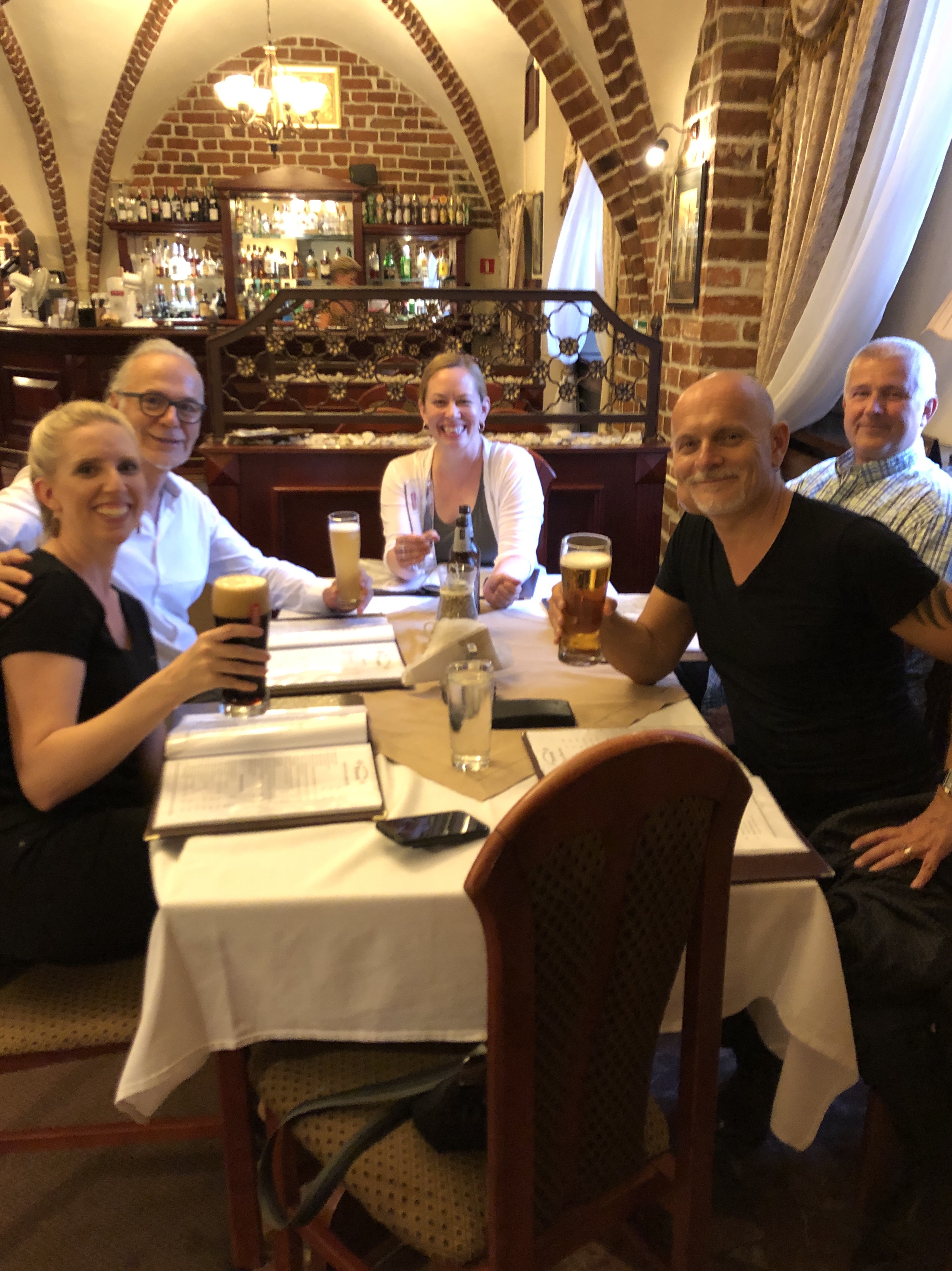
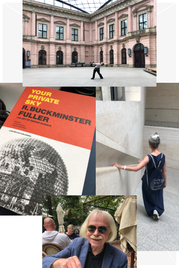
Early in my stay, there was a week-long break from working on the project, as the lab we are using was booked with another large workshop. I had planned in advance to use the time to stay with my friend and colleague, Mary Copple, who lives just outside of Berlin and shares my interest in movement and environmental space. She was able to arrange a wonderful meeting with Joachim Krausse, a Buckminster Fuller scholar. We discussed many crossovers in thoughts from science and art and discussed what makes good design elements in terms of utilizing space. In Krausse’s book about Fuller, there is a quote from Romany Marie, who worked with Fuller and made the comment, “A room should not be fixed, should not create a static mood, but should lend itself to change so that its occupants may play upon it as they would upon a piano.” In the same manner, we like to continuously challenge the fascial body system to new movement, so as not to create stagnant body patterns.
After my one-week break, I was back at the lab for two and a half more intensive weeks, which included the task of evisceration of the full body specimen and creating a window to see the fibrous pericardial sac among many other dissection projects. In the design of the plastinate, there are hints into some familiar concepts from Anatomy Trains as well as dissection elements that highlight ideas from many other fascial pioneers. I don’t want to reveal too many surprises yet, but the work is coming along very well and the project is creating a library of images as we go, that will eventually be shared.
More teams are coming into Guben during the fall and early winter to continue the work on the full body form and to create individual fascial plastinates. We are deeply grateful to all of those involved, particularly to the donors who will in a very profound sense outlast us all through their forms, helping to educate and inform us further about the fascial body.
I’ll end with a quote from anatomist Jean Fernel, who said, “Anatomy is to physiology as geography is to history; it describes the theatre of events.” All of us in this dissection project love the thrill of the endless exploration, and also of finding a story in our connected fascia that binds us together in many ways. Just as Fernel pushed forward the concept of physiology, we are all part of a new way of rethinking our anatomy. It is exciting to contribute to that process.