We’re happy to welcome back our Anatomy Trains teacher Lauri Nemetz as a guest blogger! Lauri recounts her experience presenting at this year’s Experimental Biology conference.
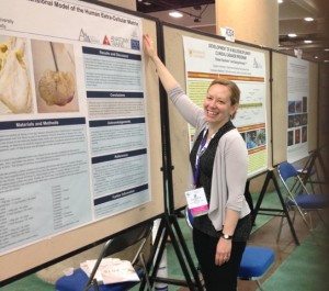
I’m back on the east coast re-settling from the Experimental Biology conference, held this year in sunny San Diego, April 2nd-5th. I am still taking in all of the information from this large conference (over 14,000 scientists and exhibitors, etc), which is a huge meeting of the minds for the biology and anatomical sciences. I feel privileged to have presented my work done in Anatomy Trains Dissection Labs as part of the American Association of Anatomists (AAA), an organization that has existed since 1888. I followed most of the conference through this track, which is a small and supportive group, with most lectures averaging around fifty people. Among my favorite sessions were “Axial Anatomy in Primates: Locomotion, Posture & Evolution” which explored early bipedalism (when we shifted to walking on two legs) and “Tissue Generation and Transplantation” which featured Luiz Sampan from the Texas Heart Institute, which had inspired my own work with fascial organs.
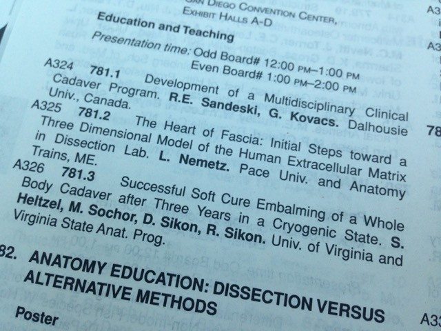
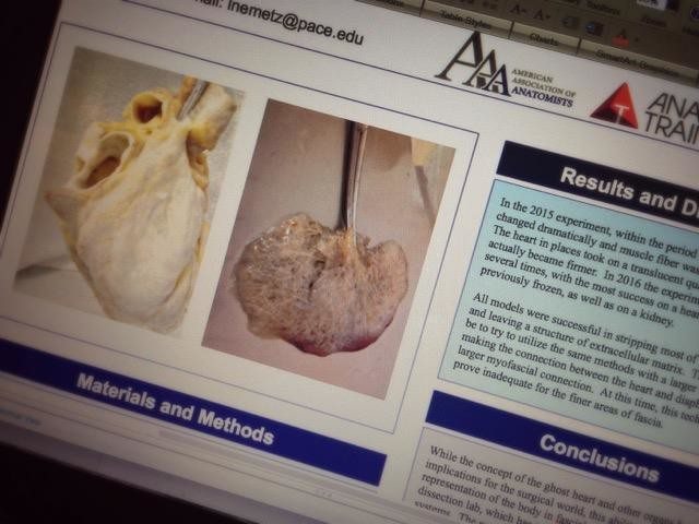
My own work and poster presentation of fascial organs garnered attention from near and far, particularly as a means to create an inexpensive fascial model in dissection lab.