From Lauri Nemetz and Tom Myers, during Fascial Dissection labs, January 2016 :

Lauri: “Traveling always gives an interesting perspective and seeing clouds, rivers, mountains, etc. echoes what we see underneath the surface of the body in lab. We have some discernible structures, and even cloud “layers” are named, but the exact beginnings and ends are really part of a larger collective whole.
Where one thing begins and another ends is more based on how we chose our perspective, while recognizing some distinct differences in composition. The question came up in class about fascia and layers. Tom expanded his answer from lab with this eloquent response”:
Tom: “The problem with defining ‘layers’ in fascia is somewhat the same as the problem in defining ‘fascia’ itself. In the middle, it’s very easy – a dissectible piece of connective fabric – but at the very small – the glycocalyx around all the cells or the endomysium around each muscle cell, e.g., too small to be dissected but clearly part of the fabric and force transmission system in the muscle. The very large is also a problem – how to term the whole fibrous network, the entire ‘adhesome’ that holds us together, maintaining a cohesive shape while necessarily allowing the shape change we call ‘movement’, as well as the shape change we call ‘developing’ or maturing.
Okay, layers: Clearly, some things have to move on other things in the body. This is obvious at the synovial joints, obvious in the organs in their ability to slide on each other, even obvious in the nerves and nerve sheaths having to slide as we stretch. We have presumed that gliding in the layers in the myofascial system is the same, with one muscle gliding over the other like two pieces of cellophane, but Gil Hedley’s Fuzz speech, Guimberteau’s films, and Huijing’s and van der Wal’s dissections and analyses have really put large holes in that theory. Muscles are connected sideways to one another and transmit force across those supposedly ‘sliding’ surfaces. (See my post on fuzz speech from last January.)
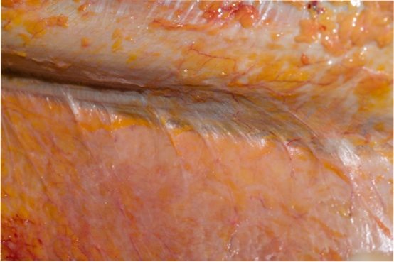
(Here’s ‘fuzz’ as it exists in an untreated cadaver. Above is the biceps, below the brachialis. It is routinely cut in dissection and rarely appears in anatomy books. It definitely allows movement between the muscles, but it also definitely limits motion and transfers force laterally between muscles. Imagine the upper muscle contracting or being stretched to the left – it would distribute force to the fascia of the lower muscle.)
When we look into muscles with ultrasound, we see a confusion of fascial planes moving up, down, and even somewhat across a muscle during contraction, putting paid to our notion of a simple sliding of filaments, all the the direction of pull of the attachments. So there is sliding within muscles, some between muscles, and maybe some between compartments. It is clear from simple experiments we have done in dissection class with acupuncture needles that different layers move in different directions during a movement (with implications for both trainers and yoga instructors) – because the different directions are somewhat dependent of the ‘driver’ for the movement – proximal or distal for instance.
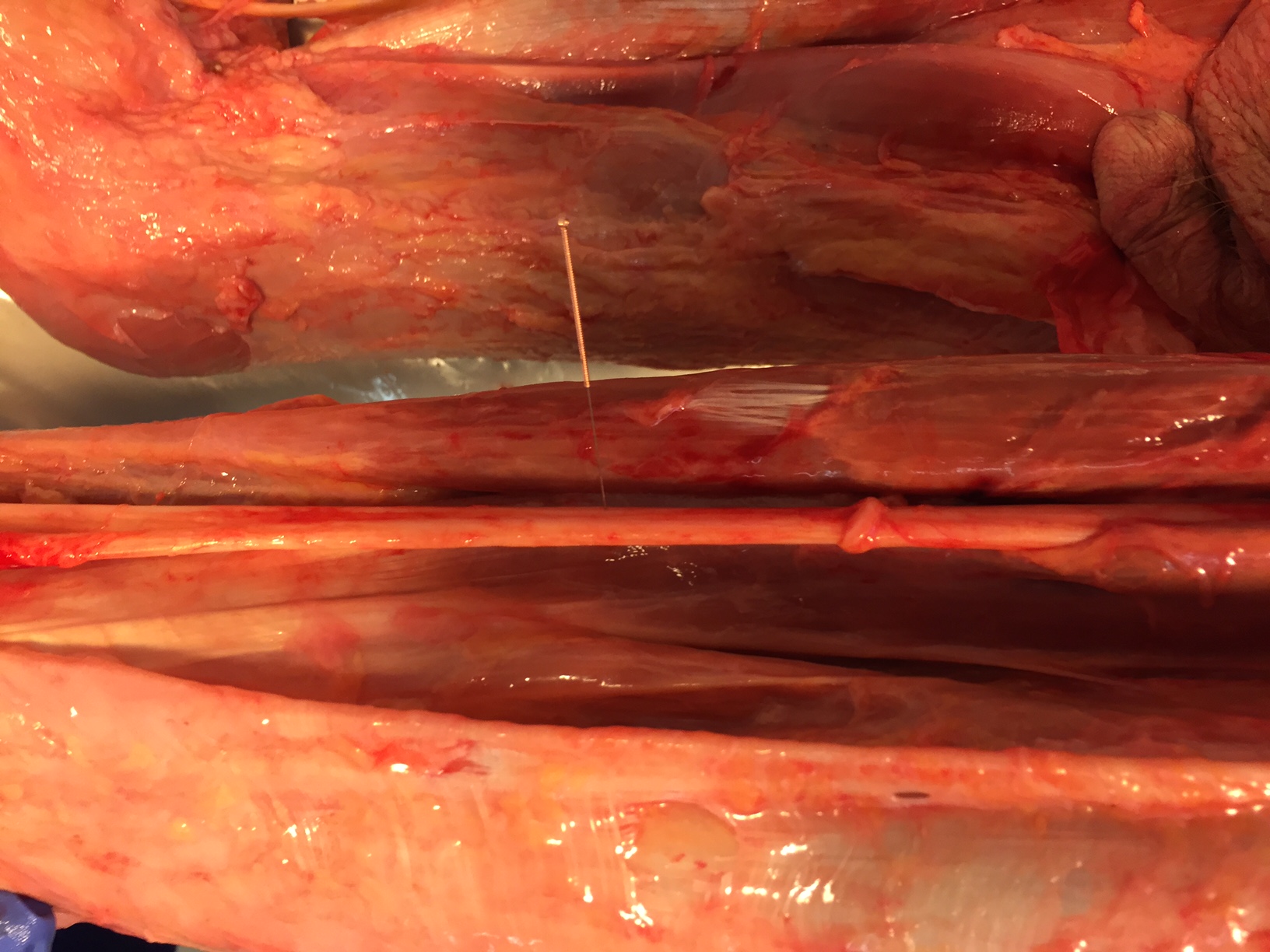
(Can you see the acupuncture needle in the muscle layers? By moving the leg we can watch the relative movement in the various fascial layers including the sheath of the sciatic nerve. Amazing to see, seeking permission to publish the video.)
But is it gliding or some form of rolling on hyaluronic foam (not foam rolling!) between tougher structures, either within or outside of a muscle. As we dissect with untreated tissue, every layer is stuck together. Stern and Stecco say the layers are rolling on the glycoprotein hyaluranon, and that when it comes in the form of very long (and maybe less then fully hydrated) chains, it is sticky. And when those hyaluranon chains are short and fully watered up, it glides, or really rolls on itself to create a gliding surface.
To cut this to the practical, two points:
- What breaks up the large and dry hyaluranon chains are all the things we have been doing: stretching, squeezing, loading, pinning and stretching, active release, and heat with movement. Keep going, this is just an updated version of the mechanism.
- Muscles clearly don’t glide on each other at the ‘origin’ end, they do at the distal end, and the middle is the juiciest and most effective place to get more differentiated movement. Let’s use the forearm’s finger flexors as an example. Up near the epicondyle, all the muscles are tied together into ‘leaves’ of fascia arising from the epicondyle, and cannot be said to slide on each other. Down at the distal end, in the carpal tunnel for instance, the tendons clearly have to slide on each other. In the middle, where the ‘muscle’ meets the ‘tendon’ (in quotes because both words are convenient fascial fictions) – that’s where stickiness happens that can be unstuck, that’s where the nervous system ‘listens’ closely with lots of GTO’s and spindles, and that’s where one can make the most difference in movement.”
Recommended
Architecture of Human Living Fascia
$74.95Architecture of Human Living Fascia summarizes 20 years of Dr. Gimberteau’s research. This widely anticipated book is richly illustrated and includes a HD DVD.
Read more